Arteries and nerves and B. Commonly called the voice box the larynx is located on top of the neck and is essential for breathing vocalizing as well as ensuring food doesnt get stuck in the trachea and cause choking.

1 5 Posterior View Of Larynx Showing Aryepiglottic And Oblique Download Scientific Diagram
In contrast to the thyroid cartilage it is a complete ring signet in shape.

. The Anatomy of the Larynx. Terminology The term commissure is a misnomer as the true vocal cords do no. Posterior View of Lord Larynx.
The epiglottis is elastic cartilage shaped like a spoon that is posterior to the root of the tongue. Well look no further. The thyrohyoid membrane was seen in the study of the neck and is pierced by the internal laryngeal nerve and superior laryngeal artery.
The inferior aspect of the. Meet the backside of Lord Larynx the producer of sound. Sitting just in front of the esophagus the vocal folds are located here making this organ absolutely.
Larynx lateral view right side. There is a printable worksheet. 950 The cartilages of the larynx.
Endoscopic view of the larynx using an office endoscope. The true vocal fold 1 extends from anterior to posterior and is separated from the false vocal fold 2 by the ventricle arrowheadsThe true folds meet anteriorly at the anterior commissure small arrow. Gemmellposts 1 Comment Although the person is speaking in German in the following video it is fun to see how of parts of your body work together to create speech and in a more sustained way singing.
Anatomy of the vocal Ligaments superior view A. Larynx anterior view The larynx is a complex hollow structure located in the anterior midline region of the neckIt is anterior to the esophagus and at the level of the third to the sixth cervical vertebrae in its normal position. Get premium high resolution news photos at Getty Images.
The posterior part of the internal space of the larynx is part of the anterior wall of the pharynx and has two vertical recesses referred to as the piriform sinus. Cartilaginous skeleton of the Larynx and Trachea A. When only air is flowing into the larynx the inlet to the larynx is open wide with the free edge of the epiglottis projecting superiorly and anteriorly.
The image is rotated 180 degrees from the usual perspective of the endoscopist. Learn vocabulary terms and more with flashcards games and other study tools. Buy this organ and its activity pages by following the links below.
This detailed 20 x 26 51 x 66 cm ENT examination-room anatomy poster depicts posterior view of the pharynx and shows sagittal section deep lateral view tonsils. The form of the lateral aspects is determined by the larynx cartilages and consist of three parts a superior one that matches the thyroid cartilage an inferior one that matches the cricoid cartilage and a middle. The Laryngeal cavity mid-sagittal view.
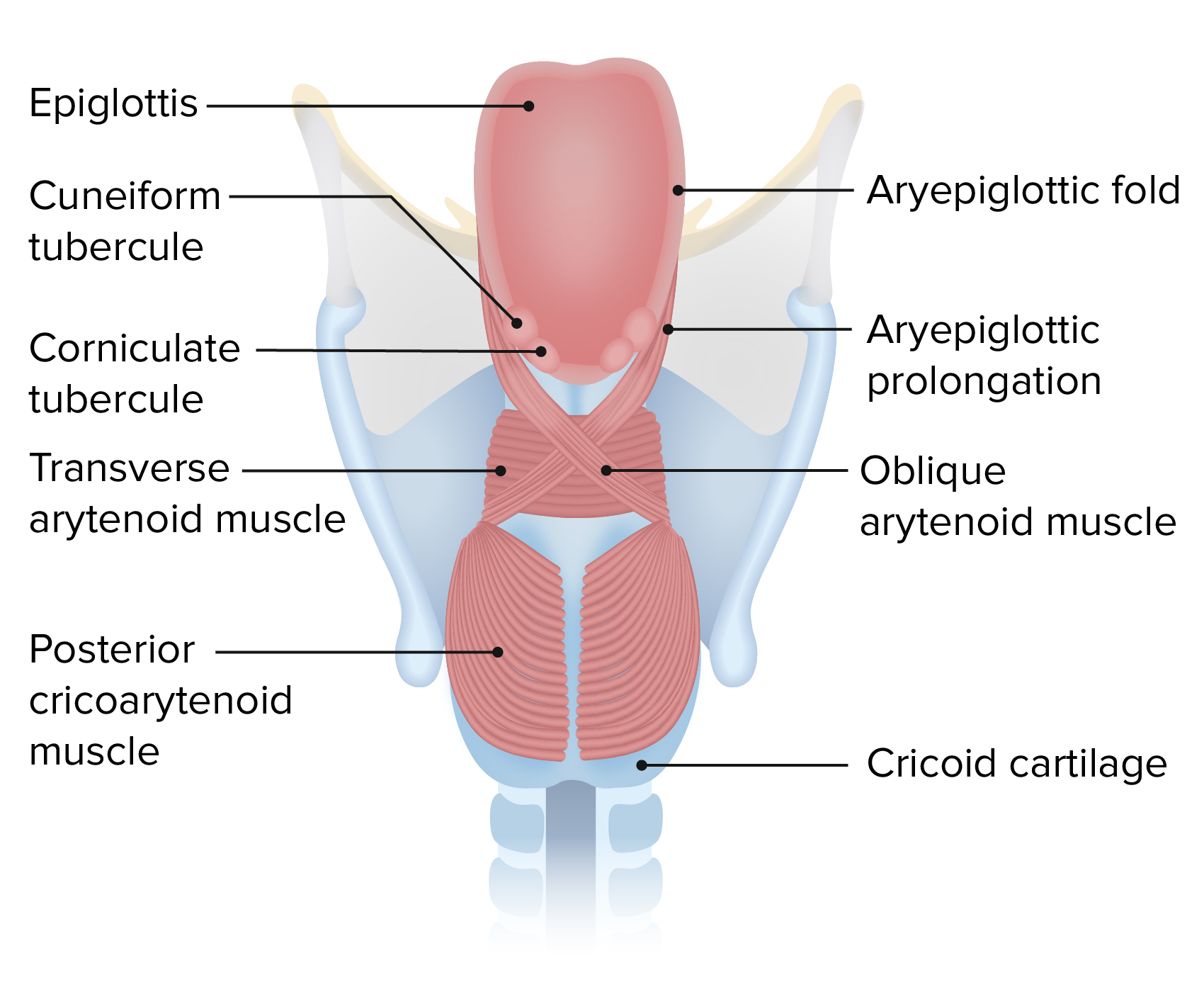
12 Posterior view of the larynx. This online quiz is called Posterior View of Larynx. Picture of Larynx and Vocal Cords Labeled Diagram stock photo images and stock photography.
A portion of the left half of the larynx above the cricoid cartilage and the muscles have been removed. This specimen comes fresh from the morgue so you can test your knowledge all the way to the exam room. Learn vocabulary terms and more with flashcards games and other study tools.
Pharynx Larynx Chart 20x26. It is situated between the trachea and the root of the tongue at the upper and. View the full answer.
Front view A epiglottis. Use the following view to review the cervical vertebrae and hyoid bone. Click on a photo for a larger view of the model.
March 3 2017 Author. Label the structures of the larynx anterior and posterior views by clicking and dragging the labels to the correct location. F upper ring of the windpipe.
Start studying Larynx Posterior View. What is larynx voice box definition where is it located anatomy cartilages muscles innervations what does the larynx do picture diagram. Posterior view of the larynx.
Part of the thyroid cartilage has been removed on the right side and the cricothyroid muscle divided. 11 and 12 The cricoid cartilage lies between the thyroid cartilage and the trachea. The posterior commissure of the larynx is a name often given to the posterior portion of the glottis.
No food or drink shall pass into his presence without dire consequences. Medial view of the right side of the larynx. Hyoid bone Epiglottic cartilage Arytenoid Cricoid cartilage Thyrohyoid ligament cartilage Thyroid cartilage Cricotracheal ligament Trachea Cricothyroid ligament Corniculate.
The larynx is contained in the visceral compartment of the neck with the thyroid gland lateral to it and the pharynx posterior to it Grants Dissector 16th ed. Various parts of the larynx area closed by connective tissue membranes. Need to osmose some anatomical knowledge.
The lower end of the epiglottis is attached to the deep surface of the thyroid cartilage. Posteriorly it has a large quadrate-shaped lamina that narrows into the arch anteriorly. Anatomy Of The Larynx Posterior View.
The larynx or organ of voice is placed at the upper part of the air passage. Lateral view and B. This high resolution full color PDF Digital Download of the larynxs posterior view is just what the doctor ordered.
Cartilages and Ligaments of the Larynx. It forms the lower part of the anterior wall of the pharynx and is covered behind by the mucous. Start studying Cartilages of the Larynx posterior view.
You need to be a group member to play the tournament. It extends from the upper border of the thyroid cartilage to the greater wing of the hyoid bone. Image Posted on September 29 2017 September 4 2018 by thecomicalanatomist.
Identify the larynx with anterior posterior lateral cut-away side views. Skull and neck osteology. 4 rows This is an online quiz called Posterior View of the Larynx.
Image LARYNX AND TRACHEA. With hemisected thyroid cartilage. The interarytenoid muscles are part of this anatomical landmark.
It consists of a cartilaginous skeleton connected by membranes ligaments and associated muscles that suspend it from. Forepart of the neck where it presents a considerable projection in the middle line. Best Way to Use This Resource.
This game is part of a tournament. Neurovasculature of the Larynx and trachea A. The thyroid cartilage has been.
The superior aspect of the cavity laryngeal inlet opens into the pharynx inferior and posterior to the tongue. Click on Label for the labeled.

Larynx Posterior View Diagram Quizlet

Muscles Of Larynx Posterior View Stock Photo Alamy

Posterior Larynx Anatomy With Annotations Wall Art Canvas Prints Framed Prints Wall Peels Great Big Canvas

Posterior View Of Larynx Human Body Vocabulary Medical Knowledge Medical Anatomy

Larynx Contemporary Health Issues
Larynx Concise Medical Knowledge
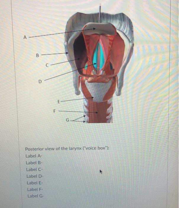
Solved A B D E F Posterior View Of The Larynx Voice Chegg Com

Cartilages Of The Larynx Posterior View Diagram Quizlet
0 comments
Post a Comment